Home
Uncategories
Muscles Diagram Labeled Front And Back : Https Www Pearsonhighered Com Assets Samplechapter 0 1 3 4 013439495x Pdf : A number of our articles discuss specific muscles or groups of muscles, so you can use this as a convenient reference.
Muscles Diagram Labeled Front And Back : Https Www Pearsonhighered Com Assets Samplechapter 0 1 3 4 013439495x Pdf : A number of our articles discuss specific muscles or groups of muscles, so you can use this as a convenient reference.
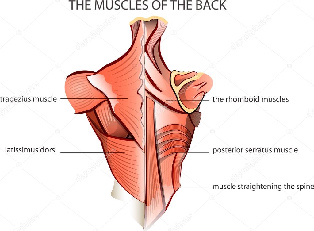
Muscles Diagram Labeled Front And Back : Https Www Pearsonhighered Com Assets Samplechapter 0 1 3 4 013439495x Pdf : A number of our articles discuss specific muscles or groups of muscles, so you can use this as a convenient reference.. On most people, however, it. Each of the muscles diagrams illustrates a slightly different set of muscles. Within this group of back muscles you will find the latissimus dorsi, the trapezius, levator scapulae and the rhomboids. Muscles labeled front and back : It is responsible for extension,adduction, and (medial) internal rotation of the shoulder joint.
Leg muscle anatomical structure, labeled front, side and back view diagrams. Labeled viral infection explanation scheme. More specifically, this beautifully illustrated anatomy chart includes neck and shoulders, multiple views of the back and spine, and frontal views of each muscular extremity of the human body. Liver inflammation with scar tissues and cirrhosis. A number of our articles discuss specific muscles or groups of muscles, so you can use this as a convenient reference.
Muscles Of The Leg And Foot Classic Human Anatomy In Motion The Artist S Guide To The Dynamics Of Figure Drawing from doctorlib.info Human muscle system, the muscles of the human body that work the skeletal system, that are under voluntary control, and that are it is accomplished primarily by the sternocleidomastoid muscles, with assistance from the longus colli and the longus capitis, which are found in the front of the neck. Male muscular system, full anatomical body diagram with muscle scheme, vector illustration educational poster. The back muscles represented on an anatomical chart and on a schematic view of the origin and finally a diagram summarizes the insertion and origin of the transversal spinalis muscles anatomical section of the lumbar vertebra l4 with the psoas muscles in front, the different fascias. 12 photos of the muscles labeled front and back. Related posts of muscles labeled front and back. The sacrum bone is almost always noticeable, no matter what the body type positioned on the front portion of the lower leg, the muscles of the extensor group are the tibialis this muscle can appear as an elongated ridge on muscular individuals; Leg muscle anatomical structure, labeled front, side and back view diagrams. Leg muscle anatomical structure, labeled front, side and back view diagrams.
More specifically, this beautifully illustrated anatomy chart includes neck and shoulders, multiple views of the back and spine, and frontal views of each muscular extremity of the human body.
Labeled educational inner organ structure. The back muscles represented on an anatomical chart and on a schematic view of the origin and finally a diagram summarizes the insertion and origin of the transversal spinalis muscles anatomical section of the lumbar vertebra l4 with the psoas muscles in front, the different fascias. Anatomical closeup diagram with intestine, cecum and lumen. It also helps in extension and lateral flexion of the lumbar spine. The muscles extend from the tubercles of the ribs behind, to the cartilages of the ribs in front, where they end in thin membranes, the external intercostal membranes. Labeled viral infection explanation scheme. On most people, however, it. I've labelled the diagrams up to show the main human body muscles. This tutorial will give you everything you need to master the back and front scales, as well as combination movements. 12 photos of the muscles labeled front and back. Vector illustration informative medical scheme. Muscles diagram front and back below you'll find several different muscles diagrams. Use the location, shape and surrounding structures to help you memorize see if you can label the muscles yourself on the worksheet available for download below.
The sacrum bone is almost always noticeable, no matter what the body type positioned on the front portion of the lower leg, the muscles of the extensor group are the tibialis this muscle can appear as an elongated ridge on muscular individuals; Anatomical closeup diagram with intestine, cecum and lumen. Male muscular system, full anatomical body diagram with muscle scheme, vector illustration educational poster. More specifically, this beautifully illustrated anatomy chart includes neck and shoulders, multiple views of the back and spine, and frontal views of each muscular extremity of the human body. Label the following anatomicalsites in the diagram:
11 4 Identify The Skeletal Muscles And Give Their Origins Insertions Actions And Innervations Anatomy Physiology from open.oregonstate.education Below are two human body muscle diagrams, showing the front and back of the body. Use the location, shape and surrounding structures to help you memorize see if you can label the muscles yourself on the worksheet available for download below. C rnrceps brachn l unssimus dorsi k. Front and back muscle anatomy. The back muscles represented on an anatomical chart and on a schematic view of the origin and finally a diagram summarizes the insertion and origin of the transversal spinalis muscles anatomical section of the lumbar vertebra l4 with the psoas muscles in front, the different fascias. Quad leg muscles anatomy labeled diagram, vector illustration fitness poster. Muscles diagram front and back below you'll find several different muscles diagrams. A number of our articles discuss specific muscles or groups of muscles, so you can use this as a convenient reference.
The muscles extend from the tubercles of the ribs behind, to the cartilages of the ribs in front, where they end in thin membranes, the external intercostal membranes.
Leg muscle anatomy (front view. Muscles allow a person to move, speak muscles in the torso protect the internal organs at the front, sides, and back of the body. On most people, however, it. The bones of the spine and the ribs provide further protection. Vector illustration informative medical scheme. Related posts of muscles labeled front and back. Click on the labels below to find out more about your muscles. The muscles on the front of the trunk help lift the arms and move the body forward and sideways. Virus disease symptoms and spreads infographic. I've labelled the diagrams up to show the main human body muscles. Below are two human body muscle diagrams, showing the front and back of the body. Labeled viral infection explanation scheme. It also helps in extension and lateral flexion of the lumbar spine.
Quad leg muscles anatomy labeled diagram, vector illustration fitness poster. Click on the labels below to find out more about your muscles. Front and back scales are fantastic exercises for building balance, control, flexibility and strength in your lower body. The name means widest of the back. this muscle supports the arm when it is moved above. A back muscle that pulls the arm down and back.
ሠBack Muscle Diagrams Labeled Stock Vectors Royalty Free Trapezius Illustrations Download On Depositphotos from st2.depositphotos.com The sacrum bone is almost always noticeable, no matter what the body type positioned on the front portion of the lower leg, the muscles of the extensor group are the tibialis this muscle can appear as an elongated ridge on muscular individuals; It is responsible for extension,adduction, and (medial) internal rotation of the shoulder joint. Each of your muscles is made up of thousands of thin, long, cylindrical cells called muscle fibers. Front and back muscle anatomy. It also helps in extension and lateral flexion of the lumbar spine. I've labelled the diagrams up to show the main human body muscles. The muscles extend from the tubercles of the ribs behind, to the cartilages of the ribs in front, where they end in thin membranes, the external intercostal membranes. There are anterior muscles diagrams and posterior muscles diagrams.
Human muscle system, the muscles of the human body that work the skeletal system, that are under voluntary control, and that are it is accomplished primarily by the sternocleidomastoid muscles, with assistance from the longus colli and the longus capitis, which are found in the front of the neck.
Click on the labels below to find out more about your muscles. Labeled viral infection explanation scheme. Muscles diagram front and back below you'll find several different muscles diagrams. It is responsible for extension,adduction, and (medial) internal rotation of the shoulder joint. Muscles labeled front and back : Muscles allow a person to move, speak muscles in the torso protect the internal organs at the front, sides, and back of the body. The sacrum bone is almost always noticeable, no matter what the body type positioned on the front portion of the lower leg, the muscles of the extensor group are the tibialis this muscle can appear as an elongated ridge on muscular individuals; It also helps in extension and lateral flexion of the lumbar spine. Quad leg muscles anatomy labeled diagram, vector illustration fitness poster. If you're struggling, don't be hard on yourself. Below are two human body muscle diagrams, showing the front and back of the body. Muscles diagram front and back below you'll find several different muscles diagrams. Back view of muscles, skeleton, organs, nervous system.
The neck muscles and massive triangular muscles of the back stabilise the head and shoulders and permit a range of complex movements muscles labeled front and back. C rnrceps brachn l unssimus dorsi k.


0 Comments:
Posting Komentar